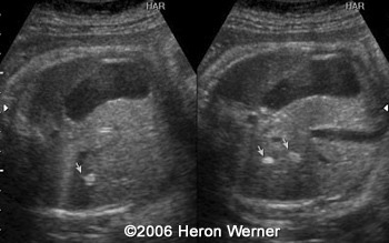
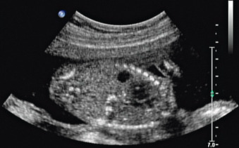
I have another scan this Tuesday and really stressed. This fetus had cytomegalovirus and was the only fetus with increasing numbers of calcifications on follow-up scans.

Fetal Intrahepatic Calcification Radiology Reference Article Radiopaedia Org
Hepatomegaly abnormal enlargement of the liver.
. The incidence of chromosomal abnormalities and genetic syndroms is not increased. Calcium deposits on babys liver detected in ultrasound. Reported that 10 of 11 cases with abnormal karyotypes had malformations that were visible on ultrasound imaging and is supported by our finding that cases without malformations are no more likely than controls without malformations to have a chromosomal abnormality 10 vs.
In most cases the reason for the calcification is never found. Staging and Treatment of Breast Cancer. I have been referred to a fetal Medicine specialist with the possibility of having an amniocentesis.
Fetal cardiac calcification is a rare ultrasound finding defined as diffuse hyperechogenicities affecting mainly the myocardium but can extend to involve the epicardium as well as the visceral pericardium. Three fetuses 21 had associated severe. The doctor is puzzled and doesnt know what this could be although he is thinking it probably isnt serious since its in the liver versus the heart brain bowels or kidneysThe ultrasound tech suggested it looked like a possible gall stone.
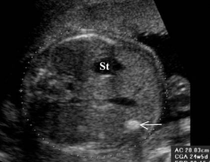
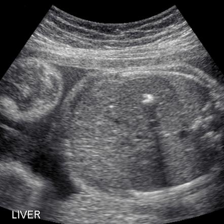
How does a Hepatic Liver Calcification happen. A hepatic calcification is an area of abnormal brightness visualised within the fetal liver. Differential of Breast Calcifications.
Im 26 weeks and really worried and confused. Characterisation of liver masses. They may be associated with aneuploidy and fetalplacental viral infections.
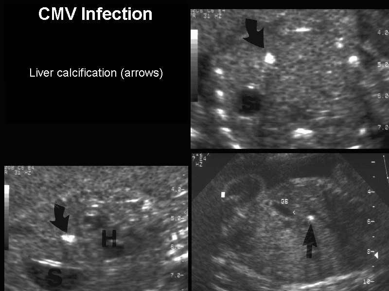
Hepatic calcifications were diagnosed in 14 fetusesan incidence of one in 1750at gestational ages of 1526 weeks. If one or more of the following signs are identified via ultrasound an amniocentesis should be done to confirm a congenital CMV infection. Liver - Segmental Anatomy.
A detailed fetal ultrasound imaging for associated abnormalities maternal STORCH syphilis cytomegalovirus herpesvirus 1 and 2 rubella and Toxoplasma analysis parvovirus serologic condition and parental cystic fibrosis mutations analysis were performed. Cases with fetal liver calcifications that were encountered between 1992 and 2001 were evaluated. The most common findings at CT are multiple low-density nodular and branching lesions up to 25 cm in diameter frequently in a subcapsular area of the liver.
Presence of solitary or multiple echogenic foci 1-2 mm in diameter within the substance of the liver or in the capsule. Outcome was determined after birth. Liver Calcifications are rare but are seen in about 1 in 1750 babies.
This fetus had cytomegalovirus and was the only fetus with increasing numbers of calcifications on follow-up scans. In most cases especially when there is only one liver calcification the baby is healthy and the. I am currently 19weeks and 2 days ago had my anatomy scan where a 7mm calcification was detected in my babys liver.
Peritoneal hepatic calcifications as calcified masses on the liver surface were reported as a feature of meconium peritonitis and. Other findings include hepatic capsule thickening subcapsular hematoma and nodular hepatic parenchymal calcifications. Our experience indicates that fetal hepatic calcification is not a rare ultrasonographic finding and each fetus with such calcifications should be thoroughly evaluated for malformations chromosomal anomalies and viral infection.
Im 26 weeks pregnant and recently had a level 2 ultrasound revealing a 4 cm spot white spot on my babys liver. Her Peri-natologist told her they could be caused by the baby swallowing blood during a bleed she had early in the pregnancy or it could be a symptom of Downs Syndrome. Four of the 33 fetuses died one of which had liver calcifications as the only sonographic finding.
Bi-RADS for Mammography and Ultrasound 2013. Calcification in the fetal liver is usually consequent to an intra-uterine infection such as cytomegalovirus toxoplasmosis rubella or syphilis. It was emphasized that the location of calcification may be affected by the causes.
It is necessary to analyse results from the. Fetal ultrasound hyperechogenities in the area of the liver can be categorized according to their location as peritoneal parenchymal and vascular. What does it mean for my baby after it is born.
Roadmap to evaluate ovarian cysts. Parenchymal calcifications may be due to intrauterine infection. Four of the 33 fetuses died one of which had liver calcifications as the only sonographic finding.
This is in line with a small study on fetal liver calcifications where Simchen et al. Twelve fetuses had one or two calcified foci one fetus had four scattered foci and one had diffuse calcification of the liver as well as peritoneal and intestinal calcifications. Typically additional ultrasound examinations are performed to make sure that the baby grows well and additional calcifications are not identified in the brain eyes abdomen or liver of the baby.
I was told that I have to be monitored every month and have a scan as my baby has calcifications on liver but they dont know what the outcome could be or wether it would be ok or not. Cases with fetal liver calcifications that were encountered between 1992 and 2001 were evaluated. The typical baby has only one calcification but some have more than one.
The purpose of this study was to provide information on the causes and postnatal outcomes of fetal liver calcifications that were detected by ultrasound imaging. Organomegaly - abnormal enlargement of organs. I have been worried and stressed since.
When isolated they are usually benign. This chapter reviews the epidemiology pathophysiology clinical and ultrasound presentation and differential diagnosis of fetal hepatic. Signs of congenital CMV infection during pregnancy.
Fetal hepatic calcifications are present in less than 1 of pregnancies. I did the TORCH test and I am still waiting for the results. My daughter is 26 weeks pregnant and two calcium deposits have been detected on the babys liver.
Outcome was determined after birth. They also said that they dont know why it has it. A detailed fetal ultrasound imaging for associated abnormalities maternal STORCH.
The calcifications can be diffuse and involve the entire myocardium or patchy over large areas of the heart. Prenatal diagnosis of liver calcifications. Thirty-three fetuses had intrahepatic calcifications at 16-38 weeks gestation.
Thirty-three fetuses had intrahepatic calcifications at 16-38 weeks gestation.
Hepatic Calcifications
Hepatic Calcifications

Hepatic Calcification Radiologic Clinics
Fetal Intrahepatic Calcification Radiology Reference Article Radiopaedia Org
Fetal Hepatic Calcification Radiology Key

Hepatic Calcification

Fetal Intrahepatic Calcification Radiology Reference Article Radiopaedia Org
Hepatic Calcification
0 comments
Post a Comment